


- Clinical Video
-
Lecture
-
Implant
Esthetic Full Arch / Denture Digital -
Surgical
Tissue Management Bone Grafting Sinus Augmentation Immediate (Placement, Loading) -
Restorative
Esthetic Materials (Ceramic, Resin) Occlusion CAD/CAM -
Digital
Guided Surgery Treatment Planning Digital Scanning (Intra Oral, Model) -
Other
Practice Management Other
-
- Case
-
Event
-
Event Conferences
International Regional -
Calendar
International Regional
-
- Publication
Search Results
Home > Search Results
Results 1 - 7 of 7 for 'CBCT'
-
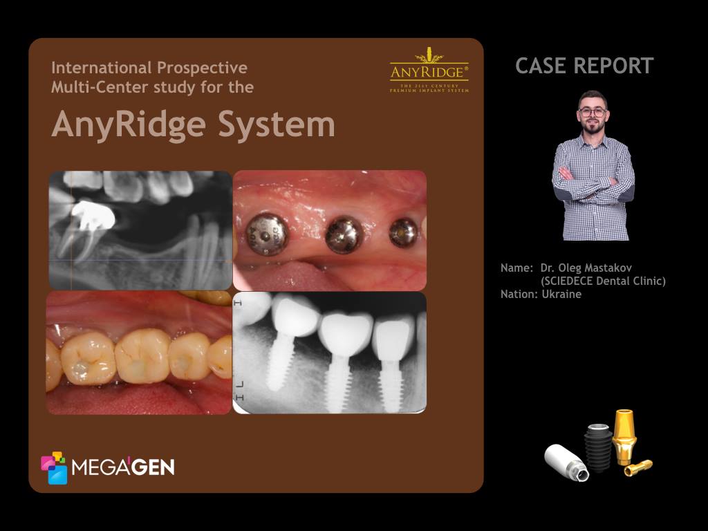
International Prospective Multi-Center s..
MegaGen UkraineClinical Case : Missing on Mandibular Right Second Premolar (#4.5) and First Molar(#4.6), Pain on the Mandibular Right Second Molar(#4.7) p.p1 {margin: 0.0px 0.0px 0.0px 0.0px; font: 12.0px Helvetica; color: #c67838; -webkit-text-stroke: #c67838} span.s1 {font-kerning: none} A 35-year-old female patient in good general health, a non-smoker was referred for consultation and treatment of 4.7 tooth with pain (Fig.3,6) after hygiene visit. Also patient expressed the desire to restore a defect of missed right second premolar 4.5 and first molar 4.6 ( Fig.1,2).There were typical narrow ridge on the mandible in position of 4.5 and 4.6 (Fig.3,5). The strategy to overcome or avoid narrow ridge problem should be considered before surgery, the diameter and the design of an implant should be considered first, according to the alveolar bone shape. Patient was against any bone grafting and healing time before implants placement, so i prefer to use an implant with small core and platform size - AnyRidge. p.p1 {margin: 0.0px 0.0px 0.0px 0.0px; font: 12.0px Helvetica; color: #c67838; -webkit-text-stroke: #c67838} span.s1 {font-kerning: none} Radiographic view of tooth 4.7 (Fig.4,6) showing a large periapical pathology and widened periodontal ligament space. Based on the clinical and radiographic findings, CBCT, it was decided to remove root 4.7 with immediate implant placement.Atraumatic root extraction technique. Before implant insertion, the dimensions of the socket were measured with a periodontal probe. As the orofacial width of the alveolus measured about 7 mm and the mesiodistal width about 7 mm, a MegaGen AnyRidge implant (diameter 4.5 mm, length 10.0 mm) was chosen.The implant was placed in an ideal three-dimensional position with high torque, the gap had to be filled with 4 APRF and wound was closed with free gingival graft and healing cap. Brain guided surgery. Placement two AnyRidge implants in position 4.5 (diameter 4.5, length 10.0 mm) and 4.6 (diameter 5.0 mm, length 10.0 mm) with high torque 70 Ncm and ISQ value 70, healing caps. Sutures 5.0 monofilament. Radiograph after surgery done (Fig.8). Three weeks after surgery photograph (Fig.7). p.p1 {margin: 0.0px 0.0px 0.0px 0.0px; font: 12.0px Helvetica; color: #c67838; -webkit-text-stroke: #c67838} p.p2 {margin: 0.0px 0.0px 0.0px 0.0px; font: 12.0px Helvetica; color: #c67838; -webkit-text-stroke: #c67838; min-height: 14.0px} span.s1 {font-kerning: none} Another 8 weeks later the healing cap was removed and an impression was taken for the fabrication of the screw retained PFM crowns. Open tray technique. Final PFM screw retained restorations photograph. (Fig 9,10)The screw retained PFM crowns constructed in lab and placed on tooth 4.7, 4.6, 4.5 with 35 Ncm torque. Access channel filled with teflon and Esthet X composite resin. Postoperative clinical photograph (Fig.11).Periapical radiograph of the implant at site 4.7, 4.6, 4.5 with crowns, one year after surgery (Fig.12).Periapical radiograph and CBCT scans of the implant at site 4.7, 4.6, 4.5 with crowns, one year follow-up after delivering the final prosthetics (Fig.13,14,15,16,17). p.p1 {margin: 0.0px 0.0px 0.0px 0.0px; font: 12.0px Helvetica; color: #c67838; -webkit-text-stroke: #c67838} span.s1 {font-kerning: none} AcknowledgmentsHygiene ProceduresDr. Dacenko Anastasia - Sciedece, Kiev, Ukraine.Surgical ProceduresDr. Mastakov Oleg - Sciedece, Kiev, Ukraine.Laboratory Procedures p.p1 {margin: 0.0px 0.0px 0.0px 0.0px; font: 12.0px Helvetica; color: #c67838; -webkit-text-stroke: #c67838} p.p2 {margin: 0.0px 0.0px 0.0px 0.0px; font: 12.0px Helvetica; color: #c67838; -webkit-text-stroke: #c67838; min-height: 14.0px} span.s1 {font-kerning: none} UA DENT lab, Kiev, Ukraine p.p1 {margin: 0.0px 0.0px 0.0px 0.0px; font: 12.0px Helvetica; color: #c67838; -webkit-text-stroke: #c67838} span.s1 {font-kerning: none} Oleg Mastakov p.p1 {margin: 0.0px 0.0px 0.0px 0.0px; font: 12.0px Helvetica; color: #c67838; -webkit-text-stroke: #c67838} span.s1 {font-kerning: none; font-variant-ligatures: no-common-ligatures} imastakov@gmail.com p.p1 {margin: 0.0px 0.0px 0.0px 0.0px; text-align: center; font: 12.0px Helvetica; color: #c67838; -webkit-text-stroke: #c67838} span.s1 {font-kerning: none} www.sciedece.com.ua p.p1 {margin: 0.0px 0.0px 0.0px 0.0px; text-align: center; font: 12.0px Helvetica; color: #c67838; -webkit-text-stroke: #c67838} span.s1 {font-kerning: none} www.facebook.com/dr.olegmastakov p.p1 {margin: 0.0px 0.0px 0.0px 0.0px; text-align: center; font: 12.0px Helvetica; color: #c67838; -webkit-text-stroke: #c67838} span.s1 {font-kerning: none} www.instagram.com/o.mastakov p.p1 {margin: 0.0px 0.0px 0.0px 0.0px; font: 12.0px Helvetica; color: #c67838; -webkit-text-stroke: #c67838} p.p2 {margin: 0.0px 0.0px 0.0px 0.0px; font: 12.0px Helvetica; color: #c67838; -webkit-text-stroke: #c67838; min-height: 14.0px} span.s1 {font-kerning: none}
Presented By : Dr. Oleg Mastakov
Hit : 1968
-

The story about one screw retained crown..
MegaGen Ukraine: The story about one screw retained crown, single AnyOne MegaGen by Dr. Oleg Mastakov, Sciedece, Kiev, UkraineA 23-year-old female patient in good general health, a non-smoker was referred for consultation and treatment of 4.7 tooth after hygiene visit. Radiographic view of tooth 4.7 showing a large periapical pathology and widened periodontal ligament space. Based on the clinical and radiographic findings, CBCT, it was decided to remove root 4.7 with immediate implant placement.Atraumatic root extraction technique. Before implant insertion, the dimensions of the socket were measured with a periodontal probe. As the oro-facial width of the alveolus measured about 8 mm and the mesio-distal width about 8 mm, a MegaGen AnyOne implant (diameter 7.0 mm, length 8.5 mm) was chosen.The implant was placed in an ideal three-dimensional position with low torque, the gap had to be filled with 6 APRF and wound was closed with free gingival graft. Sutures 5.0 monofilament.Healing cap inserted 8 weeks after implant placement for soft-tissue conditioning with ISQ implant stability probe 84/85. Another 2 weeks later the healing cap was removed and an impression was taken for the fabrication of a temporary crown with emergence profile. Acrilic crown fixed 25 N torque, vestibuloplasty done.The customized impression cap, duplicating the emergence of the restoration at site 4.7 was fabricated with flow and positioned. Open tray technique. A final A-silicone impression taken.The screw retained zirconia-supported ceramic crown constructed and placed on tooth 4.7 with 35 N torque. Access channel filled with teflon and Esthet X composite resin.Periapical radiograph of the implant at site 4.7 with crown, 3.5 months after surgery.AcknowledgmentsHygiene ProceduresDr. Nazarenko Katerina - Sciedece, Kiev, Ukraine.Surgical ProceduresDr. Mastakov Oleg - Sciedece, Kiev, Ukraine.Laboratory Procedures p.p1 {margin: 0.0px 0.0px 0.0px 0.0px; font: 11.0px Helvetica; color: #000000; -webkit-text-stroke: #000000} p.p2 {margin: 0.0px 0.0px 0.0px 0.0px; font: 11.0px Helvetica; color: #000000; -webkit-text-stroke: #000000; min-height: 13.0px} span.s1 {font-kerning: none} Yamamoto lab, Kiev, Ukraine
Presented By : Dr. Oleg Mastakov
Hit : 2902
-
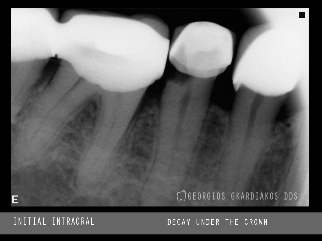
Immediate implant placement after teeth..
Immediate implant placement after teeth extractions #34 #35. At the initial intraoral xray we saw a decay under the crown of tooth #35. After clinical examination the patient also had a infection on apical area of the #34 #35. The patient was sent for a CBCT and we can see the damage of the bone near to the alveolar nerve. The treatment plan was to extract the two teeth and remove all the debris of the area. We used bone graft MEGA TCP plus A-PRF for bone regeneration. We placed the bone graft at the bottom of the socket in order to prevent contact of the implant with the alvoeral nerve. We take advantage the knife type threads and the design of the AR in order to take initial stability for the implants.
Presented By : GEORGIOS KONSTANTINOS GKARDIAKOS
Hit : 1393
-
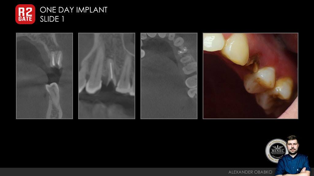
R2Gate One Day Implant Conception
- 30 years old, M, root caries #24- CBCT & Impression taking and stone model- preparation to surgery in R2GATE- #24 tooth extraction- "One Day Implant" AnyOne 3.5-11.5mm
Presented By : Aleksandr Obabko
Hit : 1250
-
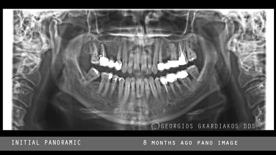
Bone Regeneration and Implant Placement
Patient came with panoramic x-ray that we could recognize bone defect at are #16 and decay on #17. The patient went for a CBCT and we saw a huge bone loss due to a apical cyst of tooth 16. Also the #17 could not be saved. We decided to extract #16 #17, remove all the debris of apical area and the granulation tissue. We make bone regeneration and wait for 8 months.After 8 months we took another CBCT to check the bone situation, then we decided to proceed to the restoration with Any Ridge implants.
Presented By : GEORGIOS K. GKARDIAKOS
Hit : 1587
-
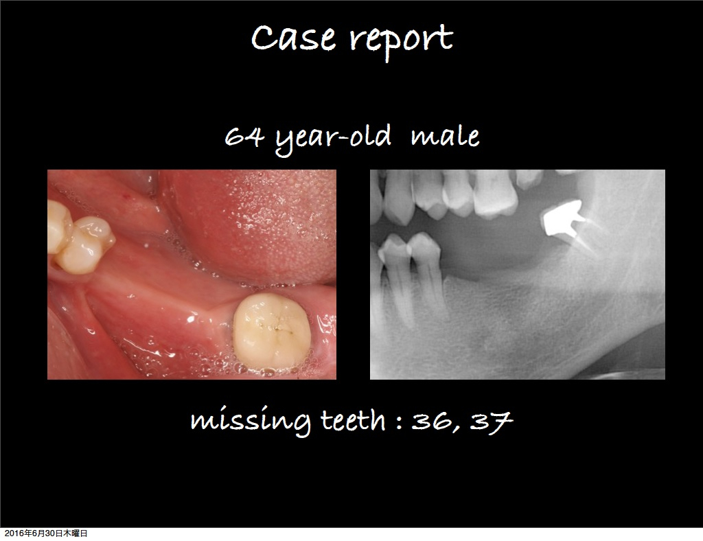
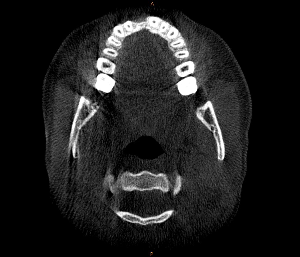
Implant Surgery with AnyRidge #36, 37 Ca..
Implant Surgery with AnyRidge #36, 37 Case Report-by Dr. Kawazoe
Presented By : Dr. Kawazoe
Hit : 2854
-
Any ridge implant placement in narrow ri..
CBCT Analysis and clinical photos
Presented By : Dr. Ahmed Mohamed El
Hit : 3270

MINEC MegaGen International Network of Education & Clinical Research
Copyright 2017 all right reserved.