


- Clinical Video
-
Lecture
-
Implant
Esthetic Full Arch / Denture Digital -
Surgical
Tissue Management Bone Grafting Sinus Augmentation Immediate (Placement, Loading) -
Restorative
Esthetic Materials (Ceramic, Resin) Occlusion CAD/CAM -
Digital
Guided Surgery Treatment Planning Digital Scanning (Intra Oral, Model) -
Other
Practice Management Other
-
- Case
-
Event
-
Event Conferences
International Regional -
Calendar
International Regional
-
- Publication
Search Results
Home > Search Results
Results 1 - 10 of 10 for 'Scan'
-
Digital Workflow: R2 Gate guided surgery..
Digital Workflow: R2 Gate guided surgery Part 2
Presented By : Miguel Stanley
Hit : 308
-

In house R2Gate surgical Guide- Digital ..
Fully Digital Workflow using R2Gate digital planning and in house surgical guide.
Presented By : Miguel Stanley
Hit : 277
-
Digital workflow for prosthetic reconstr..
The outcome of immediate implant placement can be supported by an immediate provisional restoration at the time of surgery.After the healing period the prosthetic reconstruction has to guarantee the esthetic outcome as well as the stabilization of the soft tissue contours.By using the complete digital workflow from intraoral scan, printed models and CAD CAM based final prosthetics can help to create the final restoration more easily than the conventional workflow.Thanks to the great lab support by Christopher Jehle, Zirkon Customs, Germany
Presented By : Marcus Engelschalk
Hit : 894
-
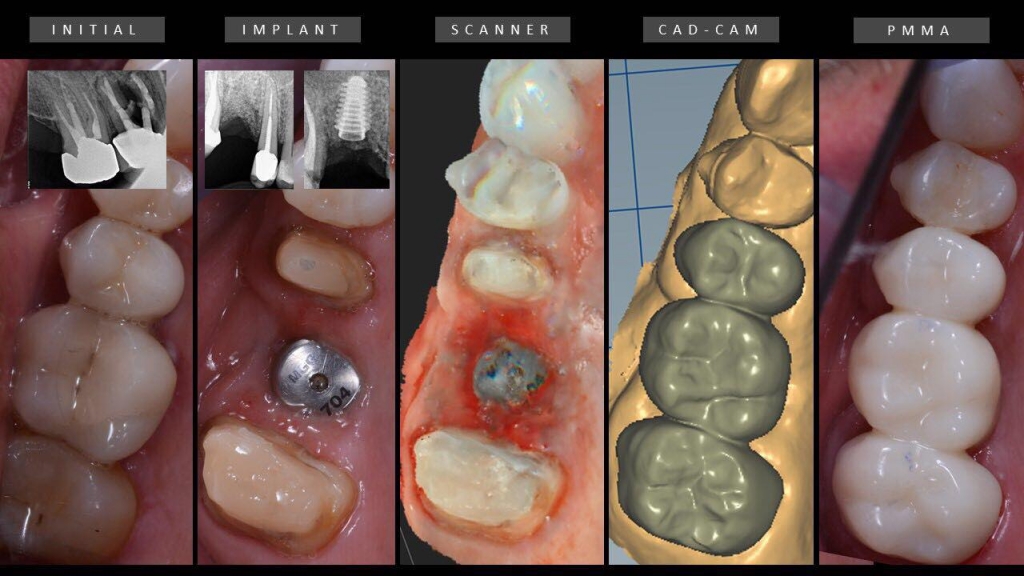
Anyridge Implant placement following a d..
A patient came to the office with a lesion on the 15. We decided to rehabilitate #15 and #16 with Megagen Anyridge implants, applying ozonetherapy, osseodensification using Versah burs, and bone regeneration applying Gen-Os bone graft mixed with i-PRF and a-PRF plug and membrane. We also followed a prosthetic digital rehabilitation protocol using the CS3600 intraoral scanner. This case is not finished yet. As you can see, a PMMA provisional bridge was placed.
Presented By : Miguel Stanley
Hit : 412
-
-
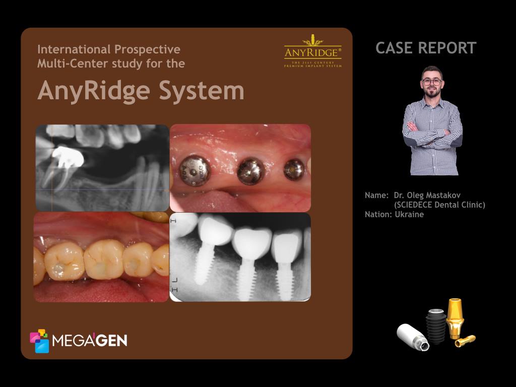
International Prospective Multi-Center s..
MegaGen UkraineClinical Case : Missing on Mandibular Right Second Premolar (#4.5) and First Molar(#4.6), Pain on the Mandibular Right Second Molar(#4.7) p.p1 {margin: 0.0px 0.0px 0.0px 0.0px; font: 12.0px Helvetica; color: #c67838; -webkit-text-stroke: #c67838} span.s1 {font-kerning: none} A 35-year-old female patient in good general health, a non-smoker was referred for consultation and treatment of 4.7 tooth with pain (Fig.3,6) after hygiene visit. Also patient expressed the desire to restore a defect of missed right second premolar 4.5 and first molar 4.6 ( Fig.1,2).There were typical narrow ridge on the mandible in position of 4.5 and 4.6 (Fig.3,5). The strategy to overcome or avoid narrow ridge problem should be considered before surgery, the diameter and the design of an implant should be considered first, according to the alveolar bone shape. Patient was against any bone grafting and healing time before implants placement, so i prefer to use an implant with small core and platform size - AnyRidge. p.p1 {margin: 0.0px 0.0px 0.0px 0.0px; font: 12.0px Helvetica; color: #c67838; -webkit-text-stroke: #c67838} span.s1 {font-kerning: none} Radiographic view of tooth 4.7 (Fig.4,6) showing a large periapical pathology and widened periodontal ligament space. Based on the clinical and radiographic findings, CBCT, it was decided to remove root 4.7 with immediate implant placement.Atraumatic root extraction technique. Before implant insertion, the dimensions of the socket were measured with a periodontal probe. As the orofacial width of the alveolus measured about 7 mm and the mesiodistal width about 7 mm, a MegaGen AnyRidge implant (diameter 4.5 mm, length 10.0 mm) was chosen.The implant was placed in an ideal three-dimensional position with high torque, the gap had to be filled with 4 APRF and wound was closed with free gingival graft and healing cap. Brain guided surgery. Placement two AnyRidge implants in position 4.5 (diameter 4.5, length 10.0 mm) and 4.6 (diameter 5.0 mm, length 10.0 mm) with high torque 70 Ncm and ISQ value 70, healing caps. Sutures 5.0 monofilament. Radiograph after surgery done (Fig.8). Three weeks after surgery photograph (Fig.7). p.p1 {margin: 0.0px 0.0px 0.0px 0.0px; font: 12.0px Helvetica; color: #c67838; -webkit-text-stroke: #c67838} p.p2 {margin: 0.0px 0.0px 0.0px 0.0px; font: 12.0px Helvetica; color: #c67838; -webkit-text-stroke: #c67838; min-height: 14.0px} span.s1 {font-kerning: none} Another 8 weeks later the healing cap was removed and an impression was taken for the fabrication of the screw retained PFM crowns. Open tray technique. Final PFM screw retained restorations photograph. (Fig 9,10)The screw retained PFM crowns constructed in lab and placed on tooth 4.7, 4.6, 4.5 with 35 Ncm torque. Access channel filled with teflon and Esthet X composite resin. Postoperative clinical photograph (Fig.11).Periapical radiograph of the implant at site 4.7, 4.6, 4.5 with crowns, one year after surgery (Fig.12).Periapical radiograph and CBCT scans of the implant at site 4.7, 4.6, 4.5 with crowns, one year follow-up after delivering the final prosthetics (Fig.13,14,15,16,17). p.p1 {margin: 0.0px 0.0px 0.0px 0.0px; font: 12.0px Helvetica; color: #c67838; -webkit-text-stroke: #c67838} span.s1 {font-kerning: none} AcknowledgmentsHygiene ProceduresDr. Dacenko Anastasia - Sciedece, Kiev, Ukraine.Surgical ProceduresDr. Mastakov Oleg - Sciedece, Kiev, Ukraine.Laboratory Procedures p.p1 {margin: 0.0px 0.0px 0.0px 0.0px; font: 12.0px Helvetica; color: #c67838; -webkit-text-stroke: #c67838} p.p2 {margin: 0.0px 0.0px 0.0px 0.0px; font: 12.0px Helvetica; color: #c67838; -webkit-text-stroke: #c67838; min-height: 14.0px} span.s1 {font-kerning: none} UA DENT lab, Kiev, Ukraine p.p1 {margin: 0.0px 0.0px 0.0px 0.0px; font: 12.0px Helvetica; color: #c67838; -webkit-text-stroke: #c67838} span.s1 {font-kerning: none} Oleg Mastakov p.p1 {margin: 0.0px 0.0px 0.0px 0.0px; font: 12.0px Helvetica; color: #c67838; -webkit-text-stroke: #c67838} span.s1 {font-kerning: none; font-variant-ligatures: no-common-ligatures} imastakov@gmail.com p.p1 {margin: 0.0px 0.0px 0.0px 0.0px; text-align: center; font: 12.0px Helvetica; color: #c67838; -webkit-text-stroke: #c67838} span.s1 {font-kerning: none} www.sciedece.com.ua p.p1 {margin: 0.0px 0.0px 0.0px 0.0px; text-align: center; font: 12.0px Helvetica; color: #c67838; -webkit-text-stroke: #c67838} span.s1 {font-kerning: none} www.facebook.com/dr.olegmastakov p.p1 {margin: 0.0px 0.0px 0.0px 0.0px; text-align: center; font: 12.0px Helvetica; color: #c67838; -webkit-text-stroke: #c67838} span.s1 {font-kerning: none} www.instagram.com/o.mastakov p.p1 {margin: 0.0px 0.0px 0.0px 0.0px; font: 12.0px Helvetica; color: #c67838; -webkit-text-stroke: #c67838} p.p2 {margin: 0.0px 0.0px 0.0px 0.0px; font: 12.0px Helvetica; color: #c67838; -webkit-text-stroke: #c67838; min-height: 14.0px} span.s1 {font-kerning: none}
Presented By : Dr. Oleg Mastakov
Hit : 1968
-

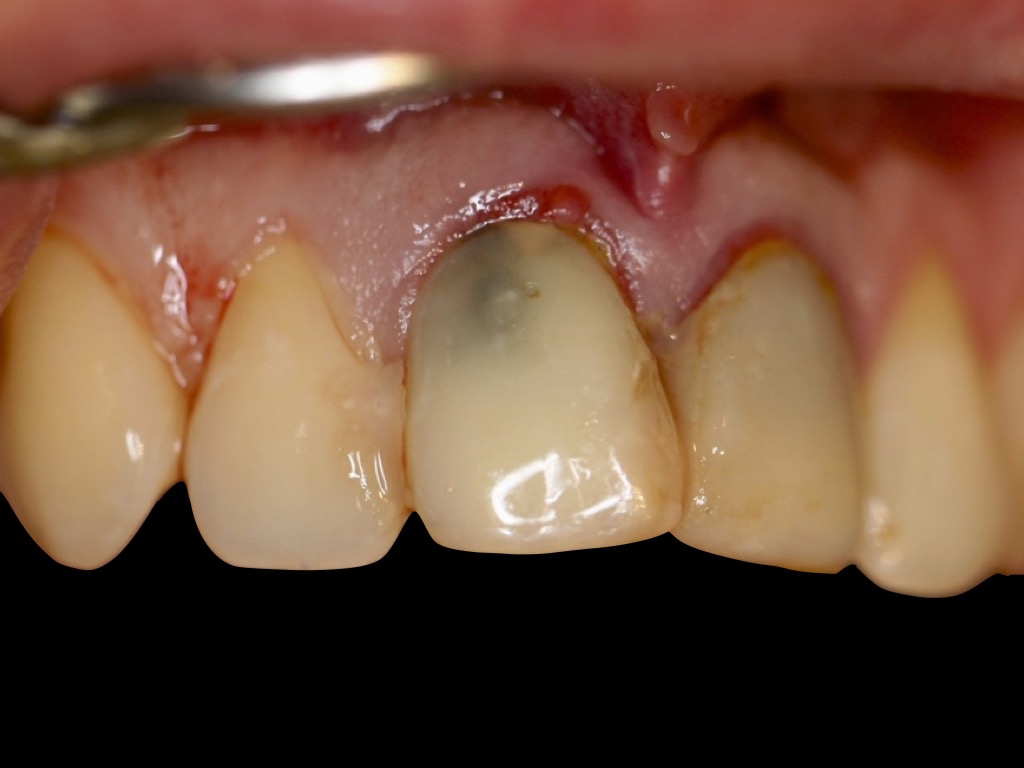
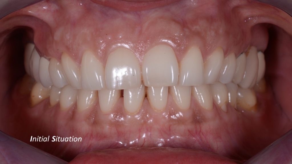
A five year follow-up on Anterior Esthet..
A 41 year old female received a root canal therapy on nº12 (US #7) after a sport accident around 2002. In october 2012 this patient come back with an history of a mountain bike accident earlier in the year. She presented with swelling, pain, probing and pus in relation to nº12 (US #7) with mesial bone loss. The diagnosis of root fracture is placed with indication of implant placement. The case is planned on the CT Scan and a surgical guide fabricated. Treatment: Immediate guided implant placement of AR, bone grafting (BG), soft tissue manipulation (STM), stock abutment placement & temporization is made (out of occlusion in all movement). This is a 5 year follow-up of nº12 (US #7) showing satisfying bone level and gingival regeneration. The ceramic crown surface was modified chair side to match the overall aspect of the rest of the arch. This is a case Classified as EI2 A-Z per the Classification developed and published regarding the EndoImplantologyTM, approach to management of endodontic challenges and complications.
Presented By : Jerome H Stroumza
Hit : 1252
-
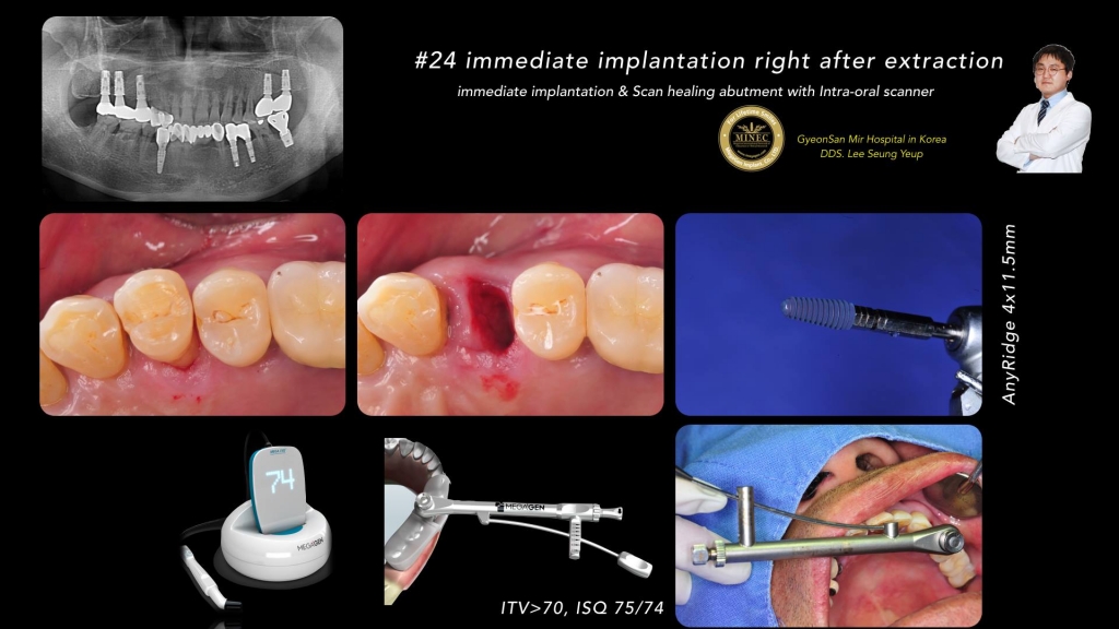
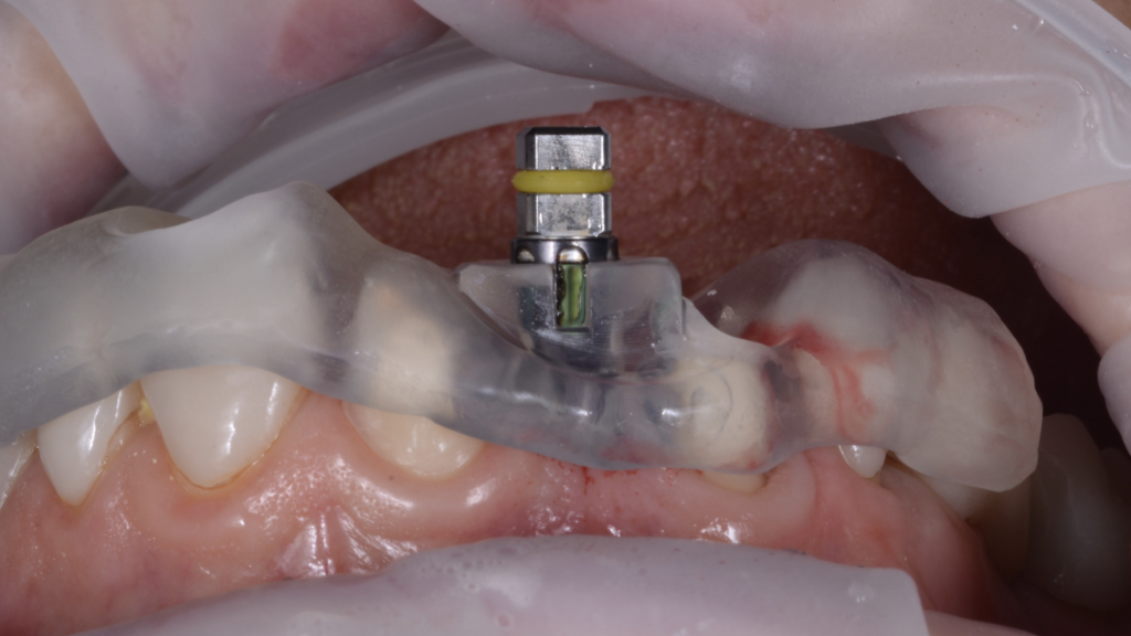
#24 Immediate Implantation right after e..
#24 Immediate Implantation right after extraction-Immediate Implantation & Scan Healing Abutment with Intra-Oral Scanner-By Dr. Seung Yeup Lee
Presented By : Dr. Seung Yeup Lee
Hit : 2035
-
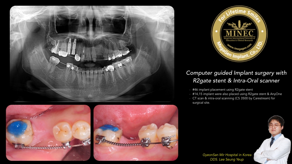
Computer guided Implant surgery with R2g..
Computer guided Implant surgery with R2gate stent & Intra-Oral scanner
Presented By : Dr. Seung Yeup Lee
Hit : 3436
-

AnyRidge Case using R2 Stent by Dr. Seun..
Case report : "Early loading with Anyridge implant using R2 Stent"- 4 Anyridge implants was placed in the lower both side.- Scan body for IntraOral scanner was connected to fixtures 3 days later after surgery.- Customized abutment & PMMA PR was delivered 10 days later after surgery.
Presented By : Dr.Seung Yeup Lee
Hit : 2107

MINEC MegaGen International Network of Education & Clinical Research
Copyright 2017 all right reserved.